CENTRAL NERVOUS SYSTEM

Development of the Central Nervous System
Following fertilisation, the nervous system begins to form in the 3rd week of development. It continues after birth and for many years into the future. Structurally, the nervous system is divided into two parts: Central nervous system – consists of the brain and the spinal cord. Peripheral nervous system – consists of cranial and spinal […]
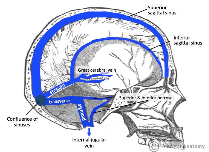
The Venous Drainage of the Central Nervous System
The central nervous system consists of the cerebrum, cerebellum, brainstem and spinal cord. Their venous drainage is complex, and rather uniquely, does not follow the arterial supply. The cerebrum, cerebellum and brainstem are drained by numerous veins, which empty into the dural venous sinuses. The spinal cord is supplied by anterior and posterior spinal veins, which drain into the […]
Structures of the Central Nervous System
[child-pages depth=”1″]
Pathways in the Central Nervous System
[child-pages depth=”1″]
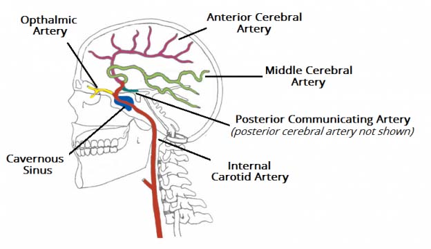
The Arterial Supply to the Central Nervous System
The central nervous system, like any system of the body, requires constant oxygenation and nourishment. The brain has a particularly high oxygen demand – at rest it represents one fifth of the body’s total oxygen consumption. It is also very sensitive to oxygen deprivation, with ischemic cell death resulting within minutes. In this article, we […]
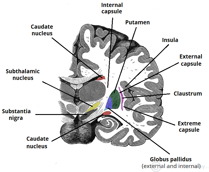
The Basal Ganglia
The basal ganglia consists of a number of subcortical nuclei. The grouping of these nuclei is related to function rather than anatomy – its components are not part of a single anatomical unit, and are spread deep within the brain. It is part of a basic feedback circuit, receiving information from several sources including the cerebral […]
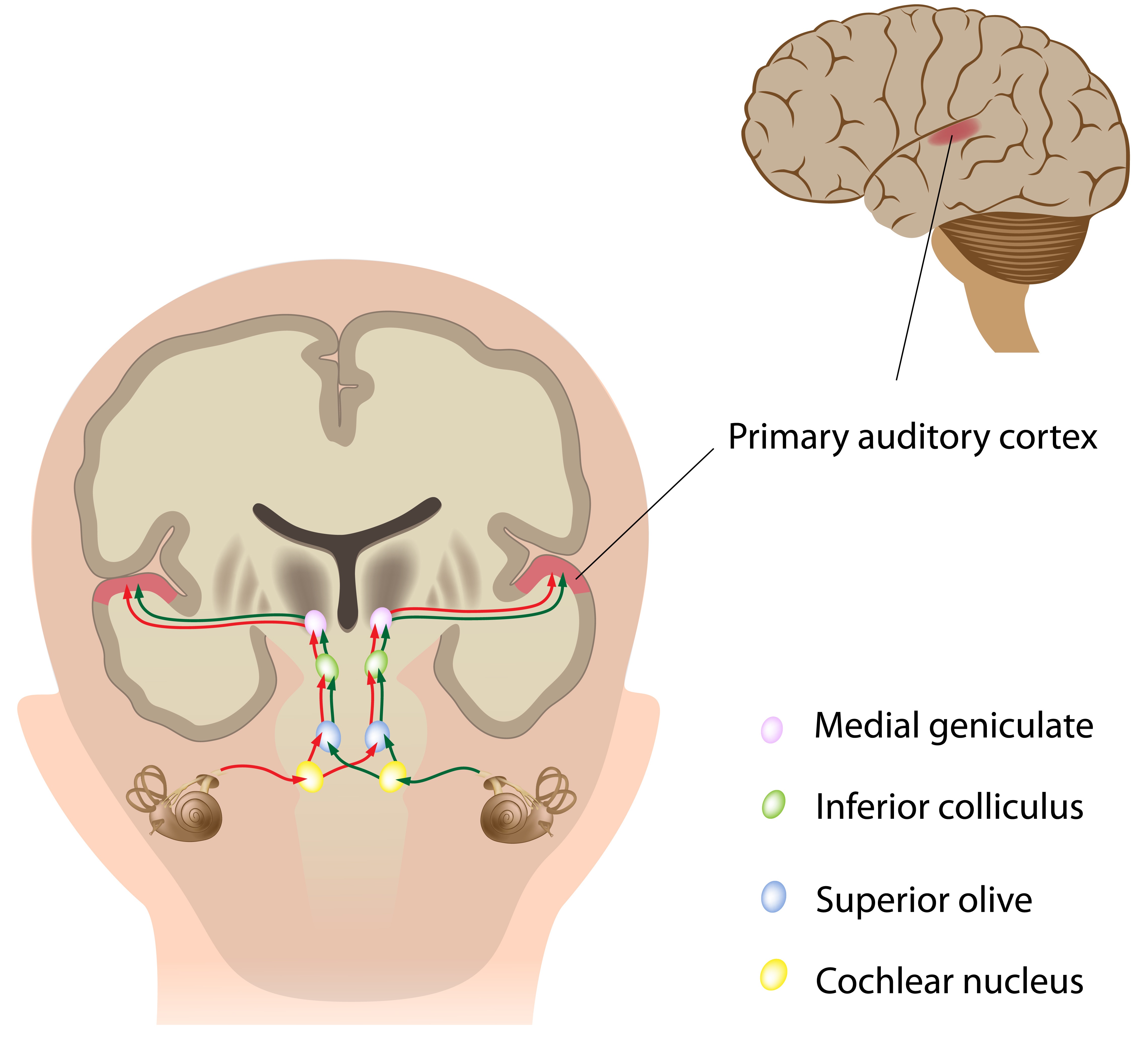
The Auditory Pathway
The auditory pathway conveys the special sense of hearing. Information travels from the receptors in the organ of Corti of the inner ear (cochlear hair cells) to the central nervous system, carried by the vestibulocochlear nerve (CN VIII). This pathway ultimately reaches the primary auditory cortex for conscious perception. In addition, unconscious processing of auditory […]

Terms of Location in Embryology
The anatomical terms of location are vital to understanding and using anatomy. They help to avoid any ambiguity that can arise when describing the location of structures. There are some terms that are specifically used in the description of embryology, which have the potential to further complicate an already complex subject! In this article, we shall look at […]
The Maxillary Division of the Trigeminal Nerve (CNV2)
The maxillary nerve is the second branch of the trigeminal nerve, which originates embryologically from the first pharyngeal arch. Its primary function is sensory supply to the mid-third of the face. In this article, we shall look at the anatomy of the maxillary nerve – its anatomical course, sensory and parasympathetic functions. Anatomical Course Trigeminal Nerve […]

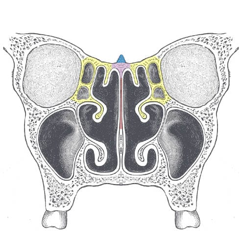
The Ethmoid Bone
The ethmoid bone is a small unpaired bone, located in the midline of the anterior cranium – the superior aspect of the skull that encloses and protects the brain. The term ‘ethmoid’ originates from the Greek ‘ethmos’, meaning sieve. This is reflected in its lightweight, spongy structure. In this article, we shall look at the anatomy […]
The ethmoid bone is a small unpaired bone, located in the midline of the anterior cranium – the superior aspect of the skull that encloses and protects the brain. The term ‘ethmoid’ originates from the Greek ‘ethmos’, meaning sieve. This is reflected in its lightweight, spongy structure. In this article, we shall look at the anatomy […]
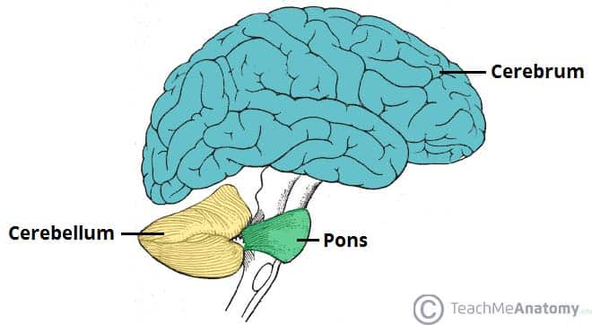
The Cerebellum
The cerebellum, which stands for “little brain”, is a structure of the central nervous system. It has an important role in motor control, with cerebellar dysfunction often presenting with motor signs. In particular, it is active in the coordination, precision and timing of movements, as well as in motor learning. During embryonic development, the anterior portion […]
The cerebellum, which stands for “little brain”, is a structure of the central nervous system. It has an important role in motor control, with cerebellar dysfunction often presenting with motor signs. In particular, it is active in the coordination, precision and timing of movements, as well as in motor learning. During embryonic development, the anterior portion […]
The Spinal Cord
The spinal cord is a tubular bundle of nervous tissue and supporting cells that extends from the brainstem to the lumbar vertebrae. Together, the spinal cord and the brain form the central nervous system. In this article, we shall examine the macroscopic anatomy of the spinal cord – its structure, membranous coverings and blood supply. […]
The spinal cord is a tubular bundle of nervous tissue and supporting cells that extends from the brainstem to the lumbar vertebrae. Together, the spinal cord and the brain form the central nervous system. In this article, we shall examine the macroscopic anatomy of the spinal cord – its structure, membranous coverings and blood supply. […]
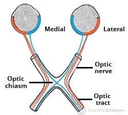
The Optic Nerve (CN II) and Visual Pathway
The optic nerve (CN II) is the second cranial nerve, responsible for transmitting the special sensory information for vision. It is developed from the optic vesicle, an outpocketing of the forebrain. The optic nerve can therefore be considered part of the central nervous system, and examination of the nerve enables an assessment of intracranial health. Due to […]
The optic nerve (CN II) is the second cranial nerve, responsible for transmitting the special sensory information for vision. It is developed from the optic vesicle, an outpocketing of the forebrain. The optic nerve can therefore be considered part of the central nervous system, and examination of the nerve enables an assessment of intracranial health. Due to […]
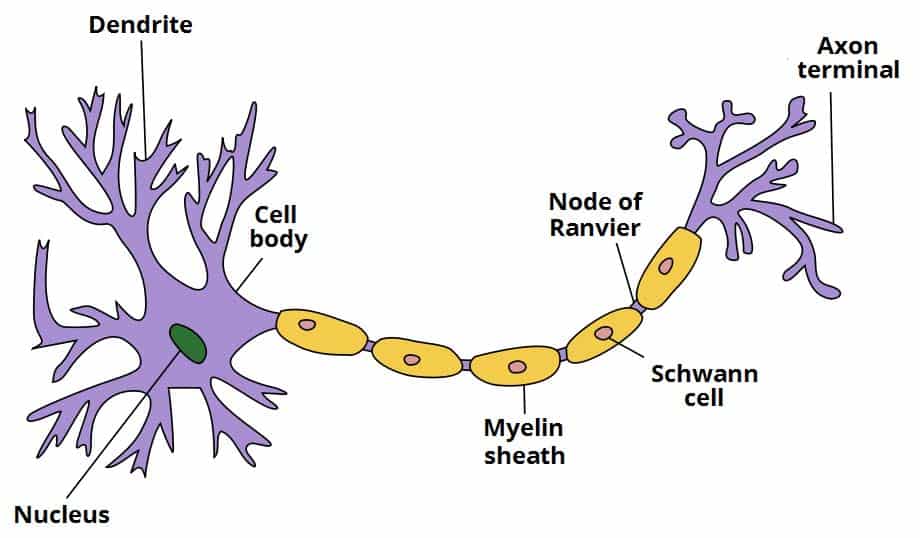
Ultrastructure of Nerves
The nervous system allows us to perceive, understand, and respond to our environment. It comprises two different types of cells: Nerve cells (neurones) – these form the functional basis of the nervous system, responsible for transmitting signals as electrical or chemical signals. Glial cells – these provide functional and structural support for the neurones. One example is […]
The nervous system allows us to perceive, understand, and respond to our environment. It comprises two different types of cells: Nerve cells (neurones) – these form the functional basis of the nervous system, responsible for transmitting signals as electrical or chemical signals. Glial cells – these provide functional and structural support for the neurones. One example is […]
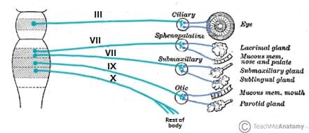
Parasympathetic Innervation to the Head and Neck
The parasympathetic nervous system is a division of the autonomic nervous system. It is involuntary, and acts with the sympathetic system to maintain body homeostasis. The actions of the parasympathetic nervous system are associated with the ‘rest and digest’ response. In this article, we shall look anatomy of the parasympathetic innervation to the head and […]
The parasympathetic nervous system is a division of the autonomic nervous system. It is involuntary, and acts with the sympathetic system to maintain body homeostasis. The actions of the parasympathetic nervous system are associated with the ‘rest and digest’ response. In this article, we shall look anatomy of the parasympathetic innervation to the head and […]
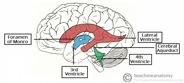
The Ventricles of the Brain
The ventricular system is a set of communicating cavities within the brain. These structures are responsible for the production, transport and removal of cerebrospinal fluid, which bathes the central nervous system. In this article, we shall look at the functions and production of cerebrospinal fluid, and the anatomy of the ventricles that contains it. Functions of Cerebrospinal Fluid […]
The ventricular system is a set of communicating cavities within the brain. These structures are responsible for the production, transport and removal of cerebrospinal fluid, which bathes the central nervous system. In this article, we shall look at the functions and production of cerebrospinal fluid, and the anatomy of the ventricles that contains it. Functions of Cerebrospinal Fluid […]
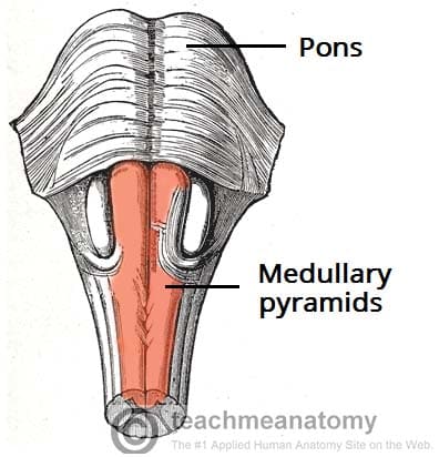
The Descending Tracts
This article is about the descending tracts of the central nervous system. The descending tracts are the pathways by which motor signals are sent from the brain to lower motor neurones. The lower motor neurones then directly innervate muscles to produce movement. The motor tracts can be functionally divided into two major groups: Pyramidal tracts – These […]
This article is about the descending tracts of the central nervous system. The descending tracts are the pathways by which motor signals are sent from the brain to lower motor neurones. The lower motor neurones then directly innervate muscles to produce movement. The motor tracts can be functionally divided into two major groups: Pyramidal tracts – These […]
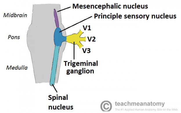
The Trigeminal Nerve (CN V)
The trigeminal nerve, CN V, is the fifth paired cranial nerve. It is also the largest cranial nerve. In this article, we shall look at the anatomical course of the nerve, and the motor, sensory and parasympathetic functions of its terminal branches. The trigeminal nerve is associated with derivatives of the 1st pharyngeal arch. Sensory: […]
The trigeminal nerve, CN V, is the fifth paired cranial nerve. It is also the largest cranial nerve. In this article, we shall look at the anatomical course of the nerve, and the motor, sensory and parasympathetic functions of its terminal branches. The trigeminal nerve is associated with derivatives of the 1st pharyngeal arch. Sensory: […]
The Appendix
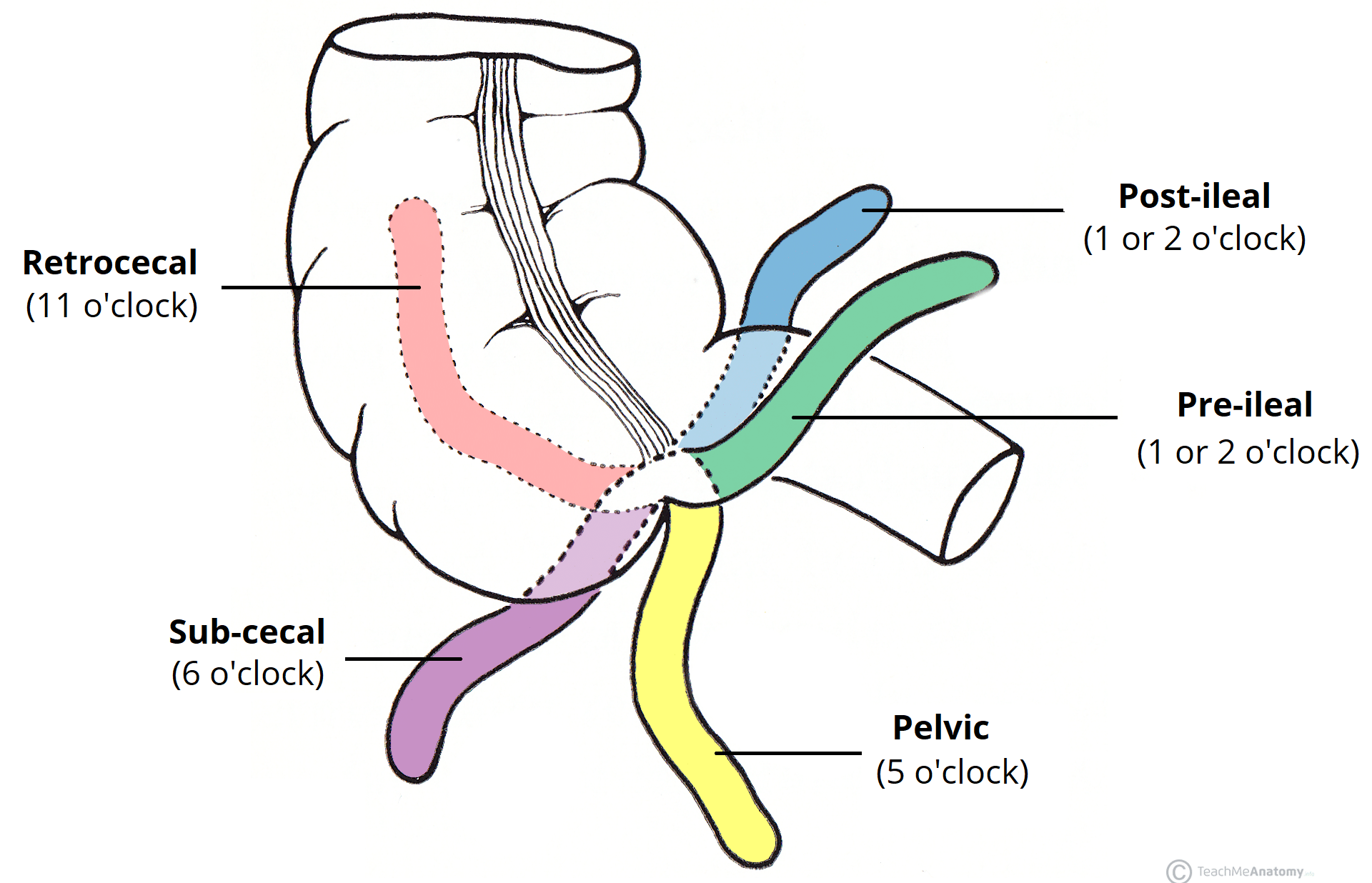
The appendix is a narrow blind-ended tube that is attached to the posteromedial end of the cecum (large intestine). It contains a large amount of lymphoid tissue but is not thought to have any vital functions in the human body. In this article, we shall look at the anatomy of the appendix – its anatomical structure and relations, neurovascular […]
The appendix is a narrow blind-ended tube that is attached to the posteromedial end of the cecum (large intestine). It contains a large amount of lymphoid tissue but is not thought to have any vital functions in the human body. In this article, we shall look at the anatomy of the appendix – its anatomical structure and relations, neurovascular […]
The Pituitary Gland
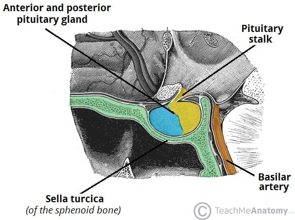
The pituitary gland (the hypophysis) is a major gland of the endocrine system. It secretes hormones that control the actions of other endocrine organs and various tissues around the body. In this article, we shall look at the anatomy of the pituitary gland – its position, structure and vascular supply. Anatomical Position and Relations The […]
The pituitary gland (the hypophysis) is a major gland of the endocrine system. It secretes hormones that control the actions of other endocrine organs and various tissues around the body. In this article, we shall look at the anatomy of the pituitary gland – its position, structure and vascular supply. Anatomical Position and Relations The […]
The Eyeball
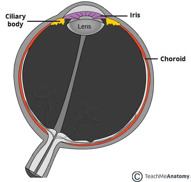
The eyeball is a bilateral and spherical organ, which houses the structures responsible for vision. It lies in a bony cavity within the facial skeleton – known as the bony orbit. Anatomically, the eyeball can be divided into three parts – the fibrous, vascular and inner layers. In this article, we shall consider the anatomy […]
The eyeball is a bilateral and spherical organ, which houses the structures responsible for vision. It lies in a bony cavity within the facial skeleton – known as the bony orbit. Anatomically, the eyeball can be divided into three parts – the fibrous, vascular and inner layers. In this article, we shall consider the anatomy […]
The Stomach
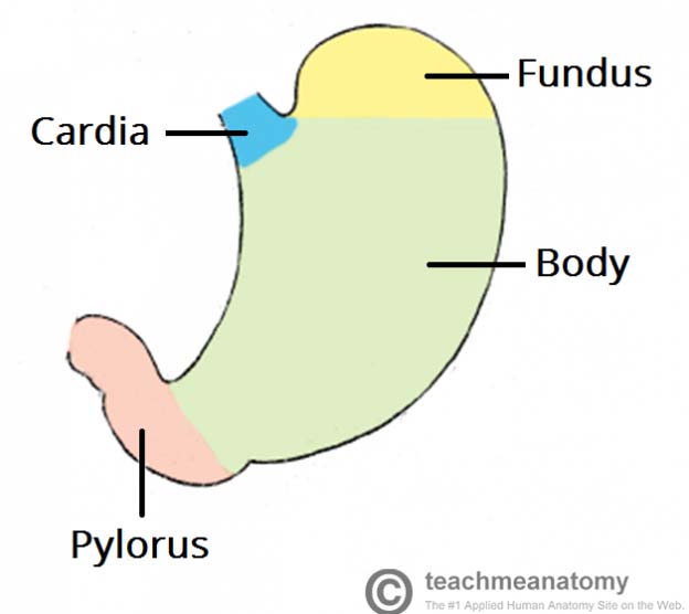
The stomach, is an intraperitoneal digestive organ located between the oesophagus and the duodenum. It has a ‘J’ shape, and features a lesser and greater curvature. The anterior and posterior surfaces are smoothly rounded with a peritoneal covering. In this article, we shall look at the anatomy of the stomach – its position, structure and neurovascular supply. Anatomical Position […]

The stomach, is an intraperitoneal digestive organ located between the oesophagus and the duodenum. It has a ‘J’ shape, and features a lesser and greater curvature. The anterior and posterior surfaces are smoothly rounded with a peritoneal covering. In this article, we shall look at the anatomy of the stomach – its position, structure and neurovascular supply. Anatomical Position […]
Ultrastructure of Muscle Cells
Muscle tissue has a unique histological appearance which enables it to carry out its function. There are three main types of muscle: Skeletal – striated muscle that is under voluntary control from the somatic nervous system. Identifying features are cylindrical cells and multiple peripheral nuclei. Cardiac – striated muscle that is found only in the heart. Identifying features are […]
Muscle tissue has a unique histological appearance which enables it to carry out its function. There are three main types of muscle: Skeletal – striated muscle that is under voluntary control from the somatic nervous system. Identifying features are cylindrical cells and multiple peripheral nuclei. Cardiac – striated muscle that is found only in the heart. Identifying features are […]


Comments
Post a Comment