GENERAL ANATOMY OF HUMAN BODY
Content -
Basic Terms and Terminology Relating to the Anatomy and Physiology of the Human Body
General Anatomy of the Human Body
Terms Relating to Anatomical Structures and Directions
Cells of the Body
Cell Processes
Tissues of the Body
Basic Terms and Terminology Relating to the Anatomy and Physiology of the Human Body
- Anatomy: The study of the parts and structures of the human body
- Physiology: The study of the functions of the human body
- Gross anatomy: The study of the parts and structures of the human body that can be seen with the naked eye and without the use of a microscope
- Microscopic anatomy: The study of the parts and structures of the human body that can NOT be seen with the naked eye and only seen with the use of a microscope
- The frontal plane: Also referred to as the coronal plane, separates the front from the back of the body.
- Ventral surface: The front of the body
- Dorsal surface: The back of the body
- Transverse plane: Also referred to as the cross sectional plane separates the top of the body at the waist from the bottom of the body
- Sagittal plane: Also referred to as the medial plane separates the right side of the body from the left side of the body
- Anterior: Closer to the front of the body than another bodily part
- Posterior: Further from the front of the body than another bodily part
- Superior: One bodily part is above another bodily part
- Inferior: One bodily part is below another bodily part
- Cytology: A subdivision of microscopic anatomy that is the study of the parts and structures of the body's cells
- Histology: A subdivision of microscopic anatomy that is the study of the parts and structures of the body's tissues
- Cell: The basic building blocks of the human body and the bodies of all other living species
- Prokaryotes: One of the two types of cells that don't have organelles or a nucleus
- Eukaryotes: One of the two types of cells that have a nucleus containing genetic material and organelles
- Cell wall: The area around the cell that protects the cell membrane and the cell from threats in its external environment
- Extracellular: The environment outside of a cell
- Intracellular: Inside the cell
- Permeability: The ability of the cell to let particles into the cells and to get particles out of the cell
- Ions: Electrically charged molecules such as electrolytes in the human body
- Anions: Electrolytes that have a negative electrical charge
- Cations: Electrolytes that have positive electrical charge
- Cell membrane: The covering that envelopes cells and somewhat acts like the gate keeper of the cell.
- Cytoplasm: The substance which makes up the bulk of a living cell and contains the organelles
- Cytoskeleton: The part of the cell that maintains the shape and form of the cell
- Cell nucleus: The place in the cell that contains chromosomes and the place where both DNA and RNA are synthesized and replicated.
- Organelles: The "mini organs" in the cell that perform a specific role.
- Mitochondria: This organelle produces and stores energy in the form of adenosine triphosphate (ATP) with a complex cycle of production known as the Krebs's cycle
- Lysosomes: The organelle that breaks down and disposes of cellular wastes
- Endoplasmic reticulum: The organelle that synthesizes proteins and lipids
- Golgi apparatus: The organelle that processes and stores the proteins and lipids that it receives from the endoplasmic reticulum
- Ribosomes: The organelle that synthesize protein with the linking of different amino acids as per the instructions of the messenger RNA molecules
- Passive transport: The movement of molecules across membranes that does NOT require the use of cellular energy to perform this transport
- Active transport: The movement of molecules across membranes that requires the use of cellular energy to perform this transport
- Diffusion: The movement of molecule from an area of higher concentration to the area or side of the membrane that has the lesser concentration
- Osmosis: A type of passive transport that does NOT require the use of cellular energy to move water and solute particles
- Meiosis: Cell division where the resulting cells have half of the original number of chromosomes
- Mitosis: Cell division where the nucleus of the cell replicates itself into two identical copies of itself
- Tissues: A group of cells with similar structure that join together to perform a specialized function
- Epithelial tissue: Also referred to as epithelium, it is the type of tissue that skin and glands are made of
- Connective tissue: The type of tissue that ligaments, tendons and bones are made of
- Skeletal muscle tissue: Striated muscle that enables voluntary bodily movement
- Smooth muscle tissue: Muscle that is not striated and not under voluntary control
- Cardiac muscle tissue: Striated, involuntary muscle that is found only in the heart. This tissue enables cardiac functioning.
- Nervous tissue: Neural tissue in the central and peripheral nervous systems
- Organs: A self-contained group of tissues that serves at least one bodily function to maintain normal bodily functioning and the homeostasis, or balance, of the body.
- Bodily systems: Groups of bodily tissues that group together to perform specific roles and functions in the body to maintain its homeostasis
General Anatomy of the Human Body
Simply stated, human anatomy is the study of the parts of the human body. Human anatomy includes both gross anatomy and microscopic anatomy. Gross anatomy includes those human structures that can be seen with the naked eye.
Gross anatomy can be compared to the structure of a house as shown in a blueprint of a house or by looking at and inspecting a house in person with the naked eye. As you look at the house's interior and exterior you will see a foundation, a roof, doors, windows, floors, a plumbing system, an electrical system, ceilings, etc. Similarly, when you view the exterior and interior of the human body with the naked eye, you are able to see its gross anatomy. For example, as you look at the human body with the naked eye, you will see its interior when the inner parts of the body are exposed, and you will see the exterior of the intact body. You will see the human's skeletal foundation, you will see the head as its roof, you will see the doors and windows in terms of the body's openings such as the mouth, the floor as the feet, an internal plumbing system with the external and internal structures and organs of the urinary and digestive systems, and you will see the brain and the heart, when exposed, as the electrical system of the body.
Microscopic anatomy, as contrasted to gross anatomy, is the study of those parts of the human body that cannot be seen with the naked eye. Structures that are viewed only with a microscope are structures included in the study of microscopic anatomy.
Microscopic anatomy is further divided into the exploration of the histological and cytological studies.
Cytology is the branch of microscopic anatomy that studies the cells and histology is the branch of microscopic anatomy that studies tissues.
From the smallest to the largest part of the human anatomy, in that sequential order, are the:
- Cells
- Tissues
- Organs
- Systems
Cells > Tissues > Organs > Systems
Terms Relating to Anatomical Structures and Directions
Anatomical Position
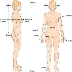
The anatomical position, with terms of relative location noted.
The anatomical position is the frame of reference for many other terms relating to anatomy, anatomical structures and anatomical directions. The anatomical position consists of a standing upright person facing forward with the person's arms on their sides next to the body and the feet together. What makes the anatomical position different from a normal standing position is the fact that the palms of the hands are unnaturally facing forward rather than naturally facing the leg, as you can see in the picture above.
Anatomical Planes
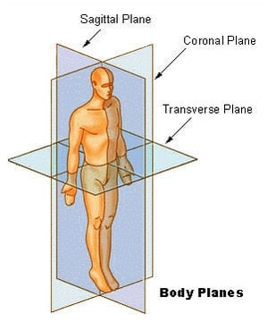
Anatomical planes in a human
Simply stated, the anatomical planes of the human body are imaginary lines going through the body that give us some point of reference when we are studying anatomy.
The frontal plane, also referred to as the coronal plane, which is shown in the picture above, is the imaginary line that separates the front from the back of the body. The term used for the front of the body is the ventral surface and the term used for the back of the body is the dorsal surface of the body.
The transverse plane, also referred to as the cross sectional plane, which is shown in the picture above, is the imaginary line that separates the top of the body at the waist from the bottom of the body.
The sagittal plane, also referred to as the medial plane, which is shown in the picture above, is the imaginary line that separates the right side of the body from the left side of the body.
Anterior and Posterior Relationships
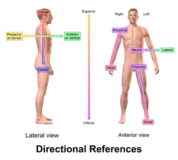
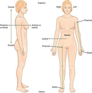
The anatomical position, with terms of relative location noted.
The term anterior is a relative and comparative directional term that is used to describe that a bodily part or anatomical structure is closer to the front of the body than another bodily part or anatomical structure. For example, the sternum, or breast bone, is anterior to the heart.
The term posterior is a relative and comparative directional term that is used to describe that a bodily part or anatomical structure is further behind another bodily part or anatomical structure. For example, the lungs are posterior to the ribs.
Superior and Inferior Relationships
- The term superior is a relative and comparative directional term that is used to describe that a bodily part or anatomical structure is above another bodily part or anatomical structure. For example, the knee is superior to the foot of the body when it is in the anatomical positon.
- Similarly, the term inferior is a relative and comparative directional term that is used to describe that a bodily part or anatomical structure is below another bodily part or anatomical structure. For example, the foot is inferior to the knee of the body when it is in the anatomical positon.
Medial and Lateral Relationships
- The term medial is a relative and comparative directional term that is used to describe that a bodily part or anatomical structure is more towards the center of the body in comparison to another bodily part or anatomical structure. For example, the nipple is medial to the shoulder.
- The term lateral is a relative and comparative directional term that is used to describe that a bodily part or anatomical structure is more away from the center of the body in comparison to another bodily part or anatomical structure. For example, the shoulder is lateral to the nipple.
Proximal and Distal Relationships
- The term proximal is a relative and comparative directional term that is used to describe that a bodily part or anatomical structure is closer to the body mass than another bodily part or anatomical structure. For example, the shoulder is proximal to the elbow.
- The term distal is a relative and comparative directional term that is used to describe that a bodily part or anatomical structure is further away from the body mass than another bodily part or anatomical structure. For example, the knee is distal to the hip.
Deep vs Superficial Relationships
- The term deep is a term to describe that a bodily part or anatomical structure is further away from the surface of the body than another bodily part or anatomical structure. For example, muscle is deeper than the skin.
- Similarly, the term superficial is a term to describe that a bodily part or anatomical structure is closer to the surface of the body than another bodily part or anatomical structure. For example, skin is the most superficial organ of the body.
Cells of the Body
Cells are the basic building blocks of the human body and the bodies of all other living species, including other mammals and plant life. Some living organisms like the amoeba and the paramecium are one celled, or unicellular, living bodies, but, for the most part, living organisms are made up of trillions and trillions of cells.
There are two different types of cells. These are prokaryotes and eukaryotes. Prokaryotes are cells that don't have organelles or a nucleus. Bacteria are an example of a prokaryote cell. Eukaryotes are cells that have a nucleus containing genetic material and organelles, as described below. The cells of the human, animals and plants are examples of eukaryote cells.
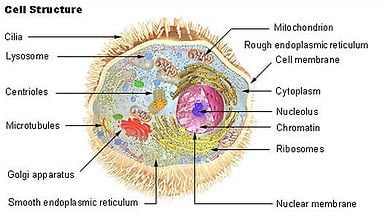
The generalized structure and molecular components of a cell
As shown in the picture above, cells consist of a:
- Cell wall
- Cell membrane
- Cytoplasm
- Cytoskeleton
- Organelles
The cell's cell wall protects the cell membrane and the cell from threats in its external environment; the external environment of the cell is referred to as extracellular. In contrast, the intracellular environment is the internal environment of the cell.
Cell membranes envelope cells and these membranes are somewhat like the gate keepers of the cell. The cell membrane performs this gate keeping function with its level of permeability. Permeability, simply defined, is the ability of the cell to let particles into the cells and to get particles out of the cell, as based on the concentration of these substances inside and outside of the cell.
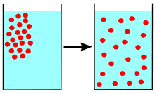
Diffusion
Diffusion is a process in physics. Some particles are dissolved in a glass of water. At first, the particles are all near one corner of the glass. If the particles randomly move around ("diffuse") in the water, they eventually become distributed randomly and uniformly from an area of high concentration to an area of low concentration, and organized.
For example, as shown above, molecules and ions can move across a cell's selective semipermeable membrane from an area of higher concentration to the area or side of the membrane that has the lesser concentration. In a sense, diffusion is the equalization of both sides of the semipermeable membrane. For example, if a substance or an electrolyte like sodium is scant in the environment outside of the cell, the semipermeable cell's membrane will release and move sodium outside of the cell to the areas of less concentration with diffusion.
Ions are electrically charged molecules such as electrolytes, in the human body. Electrolytes that have a negative electrical charge are called anions and electrolytes that have positive electrical charge are called cations. Electrolytes and the levels of electrolytes play roles that are essential to life. For example, these electrically charged ions are necessary to contract muscles, to move fluids within the body, they produce energy and they perform many other roles in the body and its physiology.
Electrolytes, similar to endocrine hormones, are produced and controlled with feedback mechanisms that control low and high levels of electrolytes.
The body's cations, or positively charged electrolytes, that move in and out of cells with diffusion, are listed below:
- Sodium which is abbreviated as Na+
- Potassium which is abbreviated as K+
- Calcium which is abbreviated as Ca+
- Magnesium which is abbreviated as Mg+
The body's anions, or negatively charged electrolytes, that move in and out of cells with diffusion, are listed below:
- Chloride which is abbreviated as Cl –
- Hydrogen phosphate which is abbreviated as HPO4–
- Bicarbonate which is abbreviated as HCO3–
- Sulfate which is abbreviated as SO4–
Cytoplasm makes up the bulk of a living cell. The major components of the cytoplasm are things like calcium, for example, the organelles which are described immediately bellow and the cytosol which makes up the bulk of a living cell. Organelles are found in the cytoplasm of the cell.
The cytoskeleton, similar to the skeletal system of the body, is made of protein and it maintains the shape and form of the cell so that it does not collapse as parts of the cell move about and the cell itself moves about.
The nucleus of the cell, as found in eurkaryotic cells, is the informational depository of the cell. The nucleus is the place that contains chromosomes and the place where both DNA and RNA are synthesized and replicated.
Organelles, which the word connotes are "mini organs" that perform a specific role in the cell. Organelles include cellular structures like the Golgi apparatus and the mitochondria, among other things, which are in the cytosol of the cell.
In addition to the mitochondria, other organelles are the:
- Lysosomes
- Endoplasmic reticulum
- Golgi apparatus
- Ribosomes
The mitochondria, as shown in the picture below, produce and store energy in the form of adenosine triphosphate (ATP) with a complex cycle of production known as the Krebs's cycle.
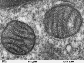
Two mitochondria from mammalian lung tissue displaying their matrix and membranes as shown by electron microscopy
Quite simplified, the mitochondria are the energy power plants of the cell.
The lysosomes, simply stated, break down and dispose of cellular wastes. Quite simplified, the lysosomes are garbage recyclers and garbage disposal systems for the cells.
Endoplasmic reticulum connects the nucleus of the cell to the cell's cytoplasm. These smooth and rough tubes and the ribosomes within play a role in the synthesis or manufacture of protein and lipids. Quite simplified, the endoplasmic reticulum can be looked at as the manufacturing plants of the cells.
The Golgi apparatus connects to the endoplasmic reticulum and it gets lipids and proteins from it. The Golgi apparatus processes these products and readies them for transport to other areas of the cell, as needed. Quite simplified, the Golgi apparatus can be viewed as the storage room for processed products.
Cell Processes
In addition to the functions and processes of the different parts of the human cell, cells also perform other processes that you should be familiar with.
These processes include:
- Passive transport
- Active transport
- Diffusion
- Osmosis
- Meiosis
- Mitosis
Passive Transport
Passive transport is the movement of molecules across membranes that does NOT require the use of cellular energy to perform this transport. Diffusion and osmosis are two forms of passive transport.
Active Transport
Active transport is the movement of molecules that does require the use of cellular energy to perform this transport.
Diffusion
Diffusion is a type of passive transport that does NOT require the use of cellular energy to move molecules, other than water molecules, from an area of higher concentration to the area of lesser concentration.
Osmosis
Osmosis is a type of passive transport that does NOT require the use of cellular energy to move water and solute particles with the stored energy found in the cell's active transport proteins.
Meiosis
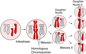
In meiosis, the chromosome or chromosomes duplicate (during interphase) and homologous chromosomes exchange genetic information (chromosomal crossover) during the first division, called meiosis I. The daughter cells divide again in meiosis II, splitting up sister chromatids to form haploid gametes. Two gametes fuse during fertilization, creating a diploid cell with a complete set of paired chromosomes. - Wikipedia
Meiosis and mitosis are two forms of cell division. When meiosis occurs, the parent or origin cell, half of the original number of chromosomes result.
Human cells have 23 types of chromosomes and each has its own set of genetic material. These 23 types of chromosomes are paired, so the human cell has a total of 46 chromosomes because 23 x 2 = 46 total chromosomes.
Meiosis consists of several phases, as shown in the picture above, that include the:
- Interphase during which time the chromosomes share and retain genetic DNA material and duplicate to create homologous chromosomes
- Meiosis I during which time the homologous chromosomes are paired up and then divided and split into two daughter nuclei
- Meiosis II during which time the two daughter nuclei divide and split into four daughter nuclei
Meiosis is the process that occurs during the fertilization of the ovum with sperm.
Mitosis
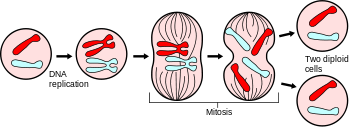
Mitosis divides the chromosomes in a cell nucleus.
Mitosis, a form of asexual replication, occurs when the nucleus of the cell replicates itself into two identical copies of itself. In other words, genetic twins result from mitosis.
The stages of mitosis in the correct sequential order, as shown in the picture above, are:
- DNA replication
- Prophase
- Prometaphase
- Metaphase
- Telophase
Tissues of the Body
Tissues are a collection or group of cells with similar structures that join to form a tissue with a distinct purpose and function. Cells collect to form tissues and tissues collect to form organs.
The four types of tissue are:
- Epithelial tissue
- Connective tissue
- Muscle tissue
- Nervous tissue
Epithelial Tissue
Also referred to as epithelium, as shown in the picture below, has several types.
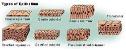
Types of epithelium
The types of epithelial tissue are:
- Columnar and shaped like columns
- Cuboidal and shaped like cubes and
- Squamous types
These different types of epithelial tissue can have one layer or they can be stratified and have multiple layers.
Epithelial tissues form all glands and they play an important role and function in the body in terms of sensations, in terms of the protection of underlying structures and organs, in terms of secretion, and in terms of absorption. It covers the entire body in the skin and it also lines the inner surfaces of organs as well as the circulatory system vessels.
The lifespan of epithelial tissue is relatively short when compared to other types of tissues, but epithelial tissue is readily replaced with mitosis cell division, as discussed above.
Connective Tissue
The type of tissue that is surrounded with what is called its matrix, as shown in the picture below:
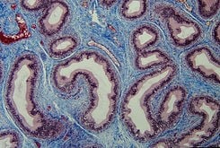
Section of epididymis. Connective tissue (blue) is seen supporting the epithelium (purple)
Connective tissue also has different types, as based on the matrix that surrounds its cells such as:
Loose Connective Tissue
Loose connective tissue lies in a soft matrix such as fluid and/or fibers. Fat, which is called adipose tissue, is an example of loose connective tissue.
Dense Connective Tissue
Dense connective tissue lies in a matrix of strong collagen fibers. Tendons and ligaments, as more fully described below in the section on the Muscular System, are comprised of dense connective tissue.
Supporting Connective Tissue
Supporting connective tissue lies in a highly firm matrix to support the body and bodily parts. Bones and cartilage are supporting connective tissues.
Fluid Connective Tissue
Fluid connective tissue lies in and is surrounded with a liquid matrix. An example of a fluid connective tissue is blood which is surrounded with plasma, the matrix for this type of connective tissue.
Muscle Tissue
As the name connotes, form the tissues of the muscles which serve to move the body.
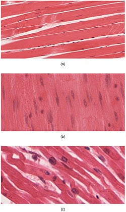
The body contains three types of muscle tissue: (a) skeletal muscle, (b) smooth muscle, and (c) cardiac muscle. (Same magnification)
As shown in the picture above there are three types of muscle tissue which are:
Skeletal Muscle
Skeletal muscle, which is also referred to striated muscle, is muscle that enables voluntary bodily movement. This tissue is composed of long muscle fibers and it is a part of all voluntary muscular movements including those used for range of motion exercises and those that serve as sphincters which control urination and defecation through the ends of the urinary and digestive systems, respectively.
Muscle fibers are stimulated or innervated by nerves to contract and relax under voluntary control.
Smooth Muscle
Smooth muscle, in sharp contrast to skeletal muscle, is not striated and it is not under voluntary control Smooth muscle is found in the digestive system where it performs peristalsis and it is also found in other organs and systems such as the vascular system where it dilates and constricts blood vessels, all of which are involuntary muscular activities.
Cardiac Muscle
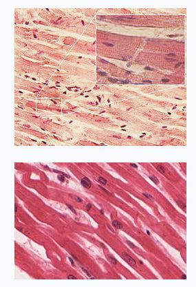
Cardiac muscle tissue, as shown in the picture above, is similar to skeletal muscle because it too is striated; however, it is also different because cardiac muscle is not voluntary like skeletal muscle is and it is not widespread throughout the body like skeletal muscle is. Cardiac muscle is restricted to the heart and it contracts and relaxes the heart during the cardiac cycle according to the electrical signals and impulses sent by the parts of the heart.
Nervous Tissue
As shown in the picture below, consists of both neural tissue and cells which are referred to as neurons:
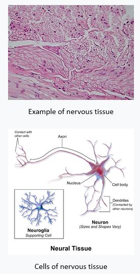
As you can see from the picture above, neurons consist of a cell body, a nucleus, an axon and dendrites that connect to other neurons.
Nervous tissue and neurons are found throughout the central nervous system and the peripheral nervous system. Simply stated, the central nervous system consists of the brain and the spinal cord, and the peripheral nervous system consists of all the other nerves and nervous tissue in the body.
The peripheral nervous system is divided into two major functional subsystems which are the:
- Autonomic nervous system
- Somatic nervous system
The autonomic nervous system controls automatic and involuntary physiological functions of the body that are outside of our control. Some of the physiological functions under the control of the autonomic, or automatic, nervous system are the movements of smooth, involuntary muscles, in contrast to voluntary skeletal muscles, like those that create peristalsis in the digestive system and the constriction of the eye's pupil when it is exposed to light.
The somatic nervous system, in sharp contrast to the autonomic nervous system, controls voluntary physiological bodily functions such as voluntary muscular movement with the skeletal muscles of the body. The somatic nervous system has efferent nerves which send and receive motor function related nerve signals and also efferent nerves which send and receive sensory function related nerve signals.




Comments
Post a Comment